Scientific challenge 3
Participate in the society

Is there an optimal size of cortical tissue pieces for cryopreservation of ovarian tissue?
Question
One important practical consideration in ovarian tissue cryopreservation (OTC) and fertility preservation is how large cortical fragments should be prepared to in order to achieve the highest survival rate of follicles after thawing and transplantation. There appear to be a considerable variation across centres, and no universal standard size. What exists are “best practices” based on balancing cryoprotectant perfusion, follicle survival, and practical surgical/transplant considerations. The aim of this letter is to discuss which considerations should be taken into account when deciding in which size to prepare cortical pieces.
Background
There is no evidence to suggest that follicle density differs systematically across ovarian geographical regions. However, follicle density may vary markedly between individual cortical fragments from the same ovary, in some cases by more than a thousand-fold (Schmidt et al., 2003). This heterogeneity probably reflects recent ovulation sites and local follicular depletion. Consequently, the number of follicles transplanted cannot be predicted based on the number of cortical pieces alone. However, when approximately half the cortical tissue from an ovary is transplanted, about half of the follicle pool will be transplanted on average.
Cortical thickness
The cortical tissue is thinned to isolate the outer cortex where almost all primordial/non-growing follicles reside and remove medulla. This is critical because follicles are located just below the ovarian surface. The widely used thickness is ≈ 1–2 mm. The rationale is that a “surface-to-volume ratio” that is sufficiently high improves penetration of cryoprotectants, ensures more uniform dehydration/cryoprotection, reduces risk of ice-crystal induced damage during freeze–thaw, and promotes better oxygen/nutrient diffusion and revascularization after transplantation.
Preparing cortical biopsies thinner than ≈1 mm (i.e., “superficial / very thin biopsies”) risks omitting primordial follicles, because follicles may lie ≈0.8 mm or more beneath the ovarian surface. Actually, some older protocols used slightly thicker pieces (e.g., up to 1.5 mm). Taken together, 1–2 mm thickness is seen as a “sweet spot” balancing good cryoprotectant perfusion and capturing the bulk of primordial follicles.
The planar dimensions (length × width) of cortical pieces
There is considerable variability in the planar dimensions of cortical pieces.
- Standard size. Cortical fragments are typically prepared as squares measuring approximately 10-5 × 5 mm with a thickness of 1 mm for the majority of adult women.
- Large ovaries (>10 mL or >10 g) If the ovary is large, cortical strips may be prepared slightly larger, typically 7–8 × 7–8 mm. Larger pieces are not preferred, as adequate tissue contact with the surrounding surface is essential to ensure rapid reperfusion after transplantation and larger pieces tend to have more difficulties being permanently situated flat on top of the underlaying tissue.
- Long cortical strips (5 × 20-15 mm). In certain cases, elongated strips are used for transplantation into the remaining ovary. These can be placed inside a subcortical tunnel with openings at each end, allowing which the tissue can to be pulled through and fixed securely in position without sutures.
- Small ovaries (children and adolescents). For small ovaries, tissue is typically prepared at 3–4 × 3–4 mm. Smaller fragments are avoided, even though follicle density is often higher in young patients, because sufficiently large pieces are needed to ensure stable positioning after transplantation with no movement of the pieces that could interfere with the establishment of new small vessels entering the tissue.
Shape of the cortical pieces
In addition, the it usually quite difficult to make pieces of similar size, often the pieces will come in many different forms and shapes depending on the size and form of the excised ovary. Square fragments are preferred because they are easier to arrange during transplantation to avoid overlap between different pieces and to allow for optimal tissue-to-tissue contact.
Below two photos to show how the pieces of cortical pieces selected for transplantation look like. As it appears the form is usually not a square and the planar dimensions of a cortical pieces is only an estimate.
Practical recommendations for cortical fragment size
Several factors influence how cortical tissue should be prepared:
- The size of the cortical tissue should be adjusted for the procurement to be performed fast and without a prolonged duration to prepare specific number or size of cortical pieces.
- The size should allow the cryopreservation procedure to be performed with optimal survival of follicles.
- The size should allow the transplantation procedure to be performed as fast as possible and with as little as possible obstacles for the pieces to be positioned in the woman.
Number of vials
For practical reasons, tissue from one ovary is normally stored in no more than 20 cryovials each containing 1 ml of cryoprotectant. If more than 20 pieces are prepared, two fragments may be placed in one vial.
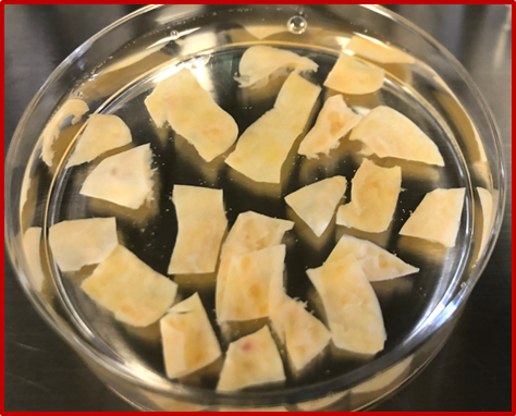
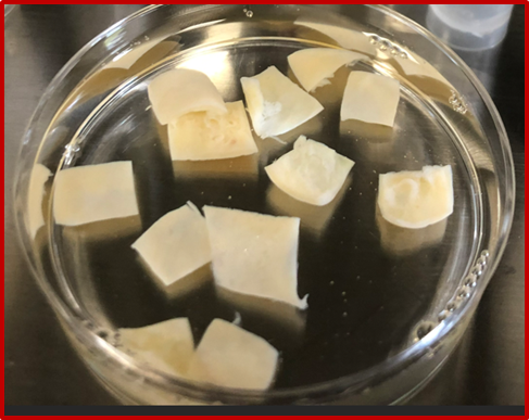
These examples of cortical strips represent the total amount of tissue to be transplanted (two patients) and does not represent all the tissue that was cryopreserved in the first place but provide an impression of how the size and shape of the tissue pieces can be prepared.
Difficulties in preparing a proper scientific investigation
There are many variables that are difficult to control, making valid answers challenging to obtain.
- The number of follicles differs largely between pieces of tissue and it will unknown how many follicles is transplanted in a given situation.
- Women in two groups needs to be comparable including similar age, diagnosis and pre-treatment AMH concentrations.
- Which end-point to use. Ideally it should be pregnancies, but will then easily take more than five years to valid data for each individual woman. AMH could be a surrogate end-point, but due to reasons listed in point 1 a large number of women should be included to get real information.
- The transplantation site should be similar between two groups.
- The actual dimensions of cortical strips are not uniform from one patients as the photos clearly indicate and how strict a selection of cortical pieces would be needed to get conclusive outcome.
Taken together, these considerations highlight the importance of discussing practices across different programs to better understand the rationale behind varying approaches to procurement and preparation of cortical fragments.
Invitation to the ISFP community
As the second ISFP Scientific Challenge, this contribution aims to stimulate open discussion on whether cortical fragment size can—or should—be further standardized, or whether flexibility based on patient, ovarian characteristics, and surgical context remains preferable. ISFP members are invited to share their protocols, experiences, observations, and views on how cortical tissue dimensions may influence cryopreservation outcomes, transplantation success, and long-term ovarian function.
References
- Schmidt KL, Byskov AG, Nyboe Andersen A, Muller J, Yding Andersen C. Density and distribution of primordial follicles in single pieces of cortex from 21 patients and in individual pieces of cortex from three entire human ovaries. Hum Reprod. 2003;18:1158-1164.
Leave a comment
You must be logged in and a member to comment.
Comments
No comments yet.